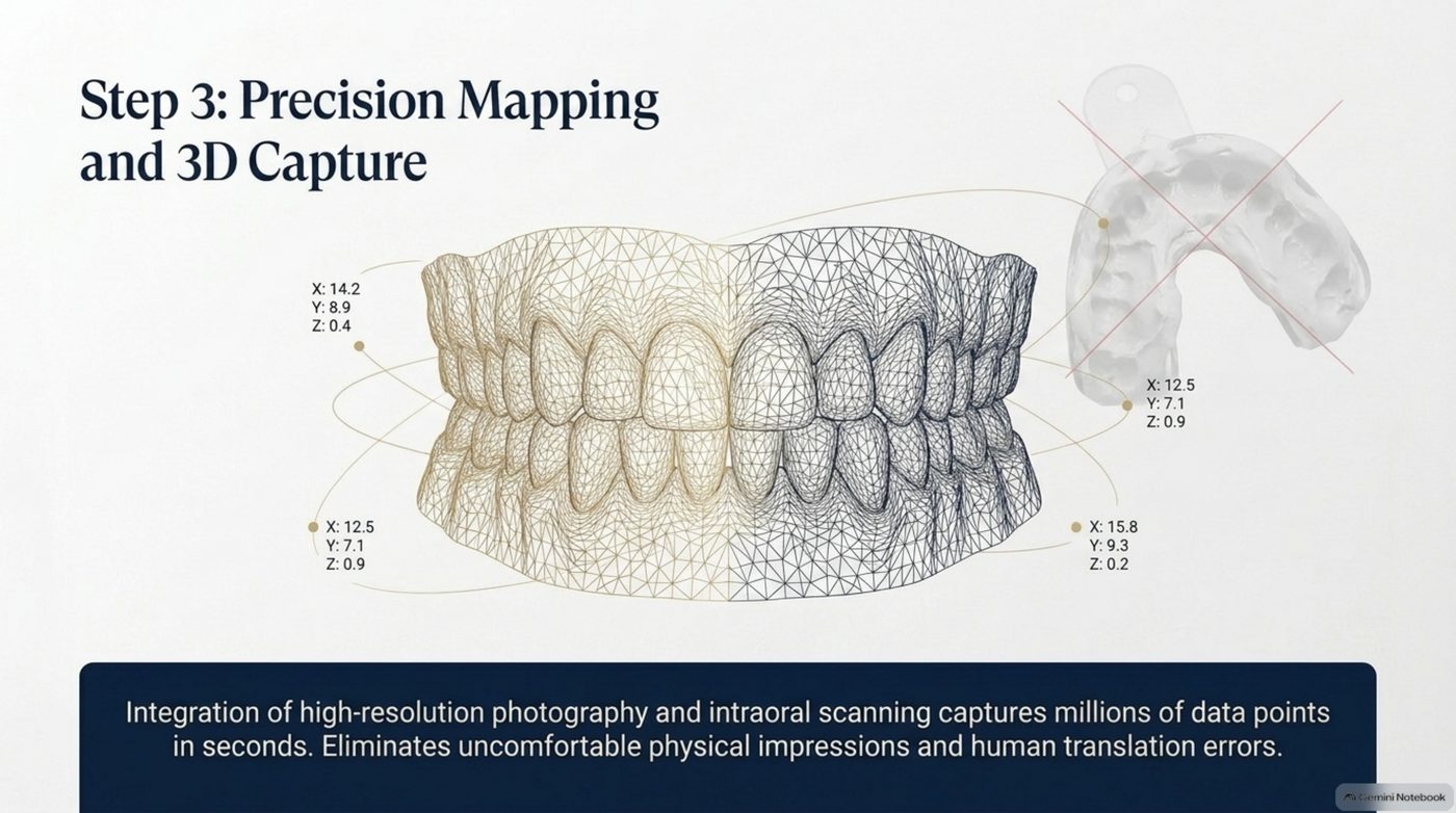
Step 3 is where the plan stops being a picture and becomes geometry. The slide behind this article shows a set of coordinate readings — X, Y and Z positions for individual landmarks — and that detail is the whole story. A smile plan that cannot be expressed in numbers cannot be fabricated precisely, and it cannot be checked against reality when it comes back from the lab. If you have been weighing up cosmetic dental treatment for a home around Sioux Falls, SD, this guide covers how it actually works, what it tends to cost, and where the results usually fall short.
Two capture technologies, one model
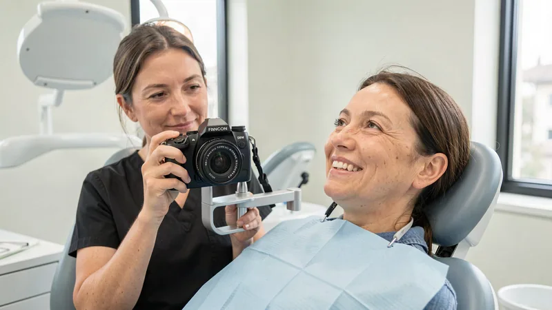
The mapping step combines two inputs. High-resolution photography records colour, translucency and surface character — the things that make a restoration read as natural rather than merely correctly shaped. Intraoral scanning records form: the exact geometry of your teeth, the contact points between them, and the relationship between the arches. Together they produce a single digital model that describes both how your teeth look and how they fit.
The deck quantifies the yield in a way that is worth pausing on: millions of data points, captured in seconds. That is not a figure of speech about thoroughness. It is the reason downstream steps can be precise — a plan built on a handful of measurements has a handful of chances to be wrong, and a plan built on millions of points has far fewer places for error to hide.
The coordinate readings, and why they matter
| Landmark reading | X | Y | Z |
|---|---|---|---|
| Position 1 | 14.2 | 8.9 | 0.4 |
| Position 2 | 12.5 | 7.1 | 0.9 |
| Position 3 | 12.5 | 7.1 | 0.9 |
| Position 4 | 15.8 | 9.3 | 0.2 |
Each row is a landmark resolved in three dimensions. The practical consequence is that a restoration can be designed, transmitted and fabricated against measured positions instead of hand-measured notes, and any deviation can be measured rather than estimated at the fitting stage.
What this step retires
The uncomfortable part of traditional capture is the physical impression: a tray of material held in place while you try not to gag, followed by a pour and a cast that can distort as it sets. Digital capture replaces the tray, and it removes a translation step as well. Every physical impression is an interpretation of your mouth; the scan is a recording of it. Removing interpretation removes a whole category of error — the kind that shows up as a restoration that is almost right.
Why it belongs before fabrication, not after
Capture that happens late can only confirm a decision; capture that happens here can make one. Because the mapping step sits between the visual confirmation and the fabrication step, the approved simulation can dictate the scan parameters and the captured geometry can drive the fabrication directly. Nothing has to be re-measured by hand in between, which is what keeps the final result aligned with the result you approved.
If you are building a picture of your own case before a consultation, the periodontal vet checklist and the smart diagnostic quiz are both free on the tools hub, and both will give you specific questions to bring to a capture appointment. To arrange mapping for your own smile, call (605) 601-8245.
Why a physical impression was never a neutral step
It is tempting to treat the impression as mere record-keeping, a neutral copy of the mouth taken before the real work begins. It is not neutral. Setting material is a chemical process that shrinks slightly and deforms as it cures, and the pour that follows is another step where the shape can shift. Add the tray seating, which is influenced by how the patient was sitting and how relaxed they were, and the result is a description of the mouth with a margin of error built into it. None of that error is anyone's fault, and it is still there in the model the technician works from. Digital capture removes the setting, the pour, and the tray from the chain.
What happens to the data afterwards
Captured geometry is only useful if it stays intact through the rest of the process. That is why this step sits in the middle of the roadmap rather than at its start. The scan is taken after the diagnostic findings have constrained the plan and after the patient has confirmed the outcome, so the data it collects is already targeted. And it is passed onward to fabrication without being re-recorded, which is what allows the geometry that reaches the machine to still be the geometry that was measured.
Colour and form are not the same problem
Mapping is sometimes described as though it were purely a shape exercise. It is not, and the roadmap's combination of photography with scanning is deliberate. Form tells the plan whether a restoration will fit and how it will meet the opposing teeth. Appearance tells the plan whether it will read as the same material as the teeth beside it. A restoration can be dimensionally flawless and still be the wrong restoration, and the only way to catch that before fabrication is to capture both.
What to ask about capture
Ask what technology will be used to record your mouth, and ask what happens to that record afterwards. A scan that is taken and then written down loses the advantage of being a scan. Ask whether the geometry passes to fabrication directly, and ask whether photographs will be captured alongside it. A plan that captures form without appearance, or that captures both and then re-measures by hand, is not a digital plan — it is a digital step inside an analog process. The smart diagnostic quiz is a useful way to arrive with specific questions rather than general ones.
Frequently Asked Questions
Is intraoral scanning uncomfortable?
It is a camera-based capture rather than a tray of setting material, so the gag reflex that physical impressions provoke is largely taken out of the process. The capture itself takes seconds rather than minutes.
Why does the plan need photography as well as a scan?
A scan records form and a photograph records appearance. Precision needs both, because a restoration that fits perfectly but reads as the wrong colour and translucency is still the wrong restoration.
Can earlier scans or photographs be reused?
Recent imaging can inform the plan, but capture for fabrication needs to describe the mouth as it is now. Teeth move, wear and change, and a plan built on outdated geometry will fabricate an outdated result.