Cosmetic dentistry has a sequencing problem. The exciting part — the visible part — is the smile design. The part that determines whether that design survives is the foundation underneath it. Step 1 of the 2026 Digital Smile Roadmap moves the foundation check to the front, and it does so with tools that see more than a human eye reading a flat image. If you have been weighing up cosmetic dental treatment for a home around Sioux Falls, SD, this guide covers how it actually works, what it tends to cost, and where the results usually fall short.
What "proactive risk planning" means
Reactive planning waits for a problem to announce itself: a cavity that has grown large enough to show on a radiograph, a tooth that has already fractured, a gum line that has already receded. Proactive planning looks for the problems that have not yet announced themselves, and it does it before the cosmetic plan is fixed. The reason is simple: a veneer placed over an undetected problem does not fix the problem, it hides it and shortens its own lifespan.
The three AI clinical findings that gate the plan
Interproximal caries detected
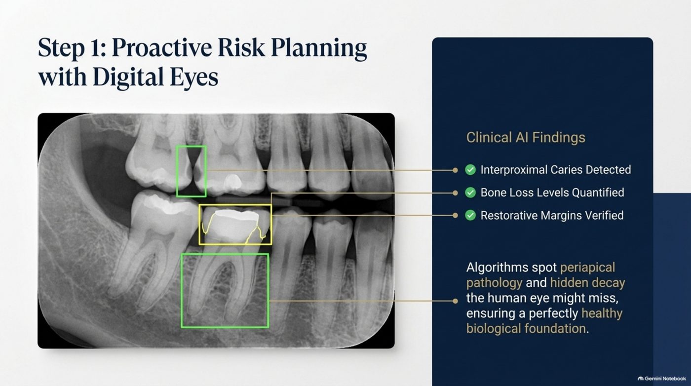
Interproximal caries sits between the teeth, in the contact areas where the surfaces are hardest to see directly and hardest to interpret on a conventional two-dimensional image. Algorithms trained on this pattern spot hidden decay that a human eye might miss, which means the roadmap can address it before a restoration is bonded over the area.
Bone loss levels quantified
Bone loss tends to be described in words — mild, moderate, advanced — and words are hard to plan around. Quantified bone loss turns that description into a measurement, which matters twice over: once for the cosmetic plan, because bone supports the gum line that frames the smile, and once for the long-term plan, because a trend is only visible if you have numbers to compare.
Restorative margins verified
Where the existing work meets the tooth is where the next problem usually starts. Verifying restorative margins before designing new work means the new restorations are built on edges that are sound, not on edges that are quietly failing.
The findings and what each one changes
| AI finding | What it quantifies | What it changes before cosmetic work |
|---|---|---|
| Interproximal caries detected | Hidden decay in contact areas between teeth | Decay is treated before a bonded restoration covers the area |
| Bone loss levels quantified | Measured bone support rather than a verbal grade | Gum-line framing and long-term trend become plannable |
| Restorative margins verified | The integrity of existing restoration edges | New work is built on verified edges, not failing ones |
What it means that algorithms spot periapical pathology
Periapical pathology develops at the tip of a tooth root, and it is the kind of finding that changes a plan completely when it is caught early and ruins a plan completely when it is not. The deck's framing is deliberately unglamorous: the purpose of detection is to ensure a perfectly healthy biological foundation. Cosmetic work is only as good as what it is attached to. A healthy foundation is not a bonus in this sequence; it is the entry condition.
How to prepare for this step as a patient
Bring your history. Recent imaging, previous restorations, any tooth that has ever been sensitive to bite pressure, and any area where flossing has changed behaviour are all useful inputs to a risk review. If you want to arrive with structure rather than notes, start with the smart diagnostic quiz and the periodontal vet checklist — both are free on the tools hub. They are not a diagnosis, and they are not a substitute for imaging, but they turn a vague worry into a specific question.
When you are ready to have the foundation assessed properly, call (605) 601-8245 to talk through what a proactive risk review would cover in your case.
The cost of catching a problem late
Late detection is expensive in a way that is not obvious from a treatment menu. A small interproximal lesion found during planning is a straightforward fix. The same lesion found after a bonded restoration has been placed over the area means the restoration has to be removed, the decay treated, and the restoration either repaired or remade — and that is the good outcome. The worse version is that the decay progresses quietly under a restoration that looks perfect from the outside, until it reaches the point where the tooth itself is at risk.
What proactive planning cannot see
No diagnostic step is complete, and it is honest to say so. Imaging sees what imaging can see, and some changes develop after the plan is set. That is why the roadmap puts the review at the front rather than treating it as a one-time gate. Findings are quantified so they can be compared later, which is what turns a single assessment into a trend. A bone level measured today and measured again in a year says something that two verbal descriptions never can.
Why the order matters more than the tools
Plenty of practices own the imaging hardware described in this step. The difference is sequencing. When risk findings arrive after the smile design is finalised, they can only force a change to a plan that has already been approved and often already begun. When they arrive first, they shape the design from the outset, which is the same information used at a point where it costs nothing to act on.
Why the foundation is the cosmetic product
It is worth stating plainly, because it is the point patients most often skip: the foundation is not preparation for the cosmetic result, it is part of it. Gum tissue frames the smile, bone supports the gum tissue, and sound margins determine whether a restoration stays sealed. A risk review is therefore not an administrative step before the interesting work. It is the step that decides whether the interesting work is still present five years from now.
Frequently Asked Questions
Does every cosmetic case need an AI risk review?
Every case benefits from a foundation check, because every restoration sits on the same teeth. The depth of the review scales with your risk profile and history rather than with the size of the cosmetic plan.
Is this step instead of, or in addition to, my regular X-rays?
In addition. The point is to extract more usable measurement from imaging you already have, so that the cosmetic plan is built on quantified findings rather than a general impression that everything looks fine.
What happens if the review finds a problem?
It moves to the front of the sequence. That is the whole purpose of putting risk planning first: the problem is addressed while it is small and before any bonded restoration depends on it.